Case Adams, PhD, Contributing Writer04.01.12
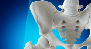
Osteoporosis afflicts more than 75 million people in the U.S., Europe and Japan; and 44 million Americans have osteoporosis, according to recent World Health Organization (WHO) and National Institutes of Health (NIH) reports.
One in two women over 60 years of age will have an osteoporosis fracture, and 10-20% with hip fractures die within six months. The 1.6 million hip fractures occurring each year in developed countries are estimated to increase to 6.3 million by 2050 as the world’s 65-and-older population grows from 524 million to 1.5 billion—and from 38 million to 85 million in the U.S.
Besides aging, generally accepted risk factors for osteoporosis include smoking, alcohol and caffeine consumption, lack of exercise and poor nutrition—all highest among developed countries.
According to Transparency Market Research, Albany, NY, the global osteoporosis drug market has grown an average of 20% per year over 2009’s $6.8 billion. Bone health supplements are also growing, but at a slower pace. According to estimates from Nutrition Business Journal (NBJ), Boulder, CO, U.S. consumer sales of bone supplements reached $1.8 billion in 2010, representing 6% growth over 2009.
According to data from SPINS, Schaumburg, IL, bone supplement sales grew 9.3% from 2010 to 2011 in combined U.S. channels. The conventional channel grew 12.4%. Vitamin D led in volume, growing 11.8%. Calcium volume followed, but grew only 4.1%. Vitamin K was last in total channel volume, but grew a hearty 23.7% in the natural channel. Magnesium sales grew 18.2% in total channel sales, and DHA sales grew 6.6%. Fish oil sales fell 1% during the year and bone-health multivitamins fell hard with a drop of 18.2%. Calcium sales in the natural channel dropped nearly 1%.
Cracks in the ‘Bricks and Mortar’
Calcium and calcium-plus-multivitamin weakness may come in part from two 2011 studies that received significant press coverage: one showing more cardiovascular events among women taking calcium; and another showing increased mortality among older women taking multivitamins.
Lackluster calcium research—showing only 16% fractures reduction after three years of 1000 mg calcium plus 400 IU vitamin D3 supplementation among 4957 volunteers—points to another issue. The “bricks and mortar” hypothesis of bone health—that bone health is achieved through bioavailability of calcium (“bricks”) along with phosphorus and other elements (“mortar”)—has some major flaws, or cracks, as it were.
An early crack getting more attention is research led by Harvard nutrition professor and leading Department of Agriculture nutritionist Dr. Mark Hegsted. Dr. Hegsted’s 1986 study, published in the Journal of Nutrition, found that countries consuming more calcium had higher hip fracture rates. The U.S. topped this list followed by New Zealand and Sweden—all top calcium-consuming countries.
Research at the University of Hong Kong in 1996 confirmed that hip fractures among the Chinese and Taiwanese were 40-50% lower than American rates.
In a 2001 review of continued research, Dr. Hegsted wrote in the American Journal of Nutrition, “Although high calcium intakes have long been recommended to prevent osteoporosis, there is little evidence that high calcium intakes effectively prevent fractures. Osteoporotic fractures are, like coronary artery disease, largely a disease of Western societies.” The data, illustrating “increasing evidence that diets high in fruit and vegetables are beneficial in preventing fractures, suggest common dietary etiologic factors.”
Confounding Bone Density
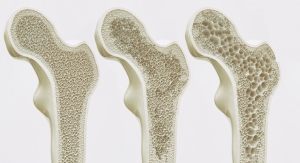
Low bone mineral density (BMD) is commonly equated with osteoporosis and bone fractures, and high BMD with health. Yet two decades of epidemiologic data indicate only about half of osteoporosis-related fracture sufferers have low bone density, or osteopenia.
A Swedish review spanning 90,000 person years and 2000 bone fractures found bone density is not an accurate indicator of an individual’s risk of fracture. “We do not recommend a program of screening menopausal women for osteoporosis by measuring bone density,” the researchers stated.
Reliance on bone density scans to gauge fracture risk is no longer acceptable. Today, the gold standard for determining fracture risk is FRAX, or Fracture Risk Assessment. FRAX factors include country of origin, clinical history, age, gender, body mass index, ethnicity, smoking, alcohol use, glucocorticoid use and rheumatoid arthritis along with osteoporosis symptoms and bone density measurements. Initially developed by WHO clinicians, FRAX is now embraced in the National Osteoporosis Foundation Clinician’s Guide and the National Osteoporosis Foundation Guide.
As far as high BMD being healthier, research published in the Journal of the American Medical Association (JAMA) found higher BMD levels linked to breast cancer. Following 6854 women over the age of 65 found breast cancer incidence 30-50% higher for every BMD unit increase.
Additionally, research by Michael Nevitt, PhD, MPH, and fellow University of California researcher Nancy Lane, MD, found high BMD levels linked to increased osteoarthritis incidence.
The Metabolic Bone
Michael McBurney, PhD, FACN, chairman of the Senior Scientific Advisory Committee at the Council for Responsible Nutrition (CRN), adjunct professor at Tufts University and head of scientific affairs at DSM Nutritional Products North America, Parsippany, NJ, clarified the nature of bones. “Despite their apparent rigidity, bones are metabolically active,” he noted. “Bone constantly remodels through bone resorption mainly by osteoclasts, and bone formation mainly by osteoblasts. As people age, bones may become brittle or osteoporotic when osteoclast activity exceeds osteoblast activity.”
“Metabolically active” departs from the “bricks and mortar” thesis, but minerals are still the major component. “Bone,” said Dr. McBurney, “is composed of minerals (50-70%), an organic matrix (20-40%), water and lipid.”
“Bones are a living substance and calcium is one important building element,” added Mona Møller, PhD, chief science officer of Norway-based Kappa Bioscience AS. “Bone mass increases until the early 20s, when it starts to decline. After 30 years of age, bone mass is decreasing in both men and women.”
A New Sheriff in Town
In 2005, researchers at Korea’s University of Ulsan College of Medicine found osteopenia and osteoporosis linked to 54% and 35% higher C-reactive protein (CRP) levels, respectively, among 4693 pre- and postmenopausal women. They also found significantly higher total alkaline phosphatase (ALP).
Both markers—CRP and ALP—indicate systemic inflammation. They are prevalent in cardiovascular disease and other inflammatory disorders. “These findings suggest that subclinical systemic inflammation may be associated with bone turnover rate and bone mass in healthy women,” the researchers stated.
University of Aberdeen Medical School researchers found the pro-inflammatory nitric oxide synthase pathway activated in osteoporosis. Sufferers had up to 344% higher nitric oxide levels, and higher pro-inflammatory nitric oxide levels yielded 64% lower bone density than controls.
Anthony Yun, MD, and Patrick Lee, MD, from Stanford University have linked osteoporosis to inflammation, and a flurry of research has confirmed the hypothesis. University studies from around the world have established that low BMD and osteoporosis are associated with pro-inflammatory mediators IL-1β, IL-6, TNF-α, as well as RANK/RANKL.
University of Zurich researchers characterized osteoporosis as relating to “systemic inflammation, oxidative stress, hypoxia and sympathetic activation.”
University of Maryland research led by Natalie Silverman, MD, found aerobic exercise decreased bone loss together with inflammation markers IL-6 and TNF-α among 86 postmenopausal overweight women.
The Sheriff’s Deputies
Research has also been pinpointing the mechanisms. Last year, Scranton Commonwealth Medical College research confirmed bone’s remodeling processes are regulated by an extracellular protein called connective tissue growth factor (CTGF).
CTGF regulates the building of blood vessel walls, joints and other tissues, as well as the repair and regeneration of bone matrix by osteoblasts. CTGF uses an immune system signaling cytokine called TGF-β1. TGF-β1 is expressed by regulating T-cells (rT-cells), which also regulate inflammatory wound repair.
Yet over-expressed CTGF (and TGF-β) drives bone turnover overboard, inhibiting bone morphogenetic proteins (BMPs) that mitigate bone loss.
BMP7, however, can shut down CTGF expression, halting TGF-β1 and bone loss. BMP7 rearranges priorities between mesenchymal stem cells, osteoblasts and osteoclasts, encouraging bone growth.
University of Amsterdam researchers found BMP7 expressed in mechanical loading, connecting weight-bearing to increased bone strength. Vitamin D apparently stimulates BMP7 as well.
BMPs regulate the matrix metalloproteinase (MMP) enzymes produced by osteoclasts and osteoblasts. These govern bone strength. BMP7s are also expressed in cancer, and they inhibit vascular calcification, or hardening of the arteries.
TGF-β1 and NF-kB are the key mediators in bone loss. While T-cell-produced TGF-β1 regulates bone remodeling, NF-kB regulates inflammatory bone loss at the genetic level. NF-kB can be stimulated by TNF-α, IL-1 and others. Meanwhile, TGF-β1 modulates IL-1, IL-2 receptors, IL-6 and others. These countervailing inflammatory mediators—NF-kB and TGF-β1—provide key mechanisms linking bone loss to inflammation.
Illustrating this notion, German researchers isolated osteoblasts from femurs of patients undergoing total hip replacement. TGF-β1 mediators proliferated among the osteoblasts. They reduced osteocalcin, increased osteopontin and decreased BMPs, accelerating bone loss.
Vanderbilt University researchers found TGF-β1 increases parathyroid hormone-related protein (PTHrP) activity by stimulating the Gli2 signaling molecule, speeding bone loss.
In other words, bone loss is an inflammatory event.
Implications of Inflammatory Bone Loss
Harvard School of Public Health researcher Dallas Jones, PhD, has characterized the study of inflammatory bone loss as “osteoimmunology.” University of Lille Nord de France researchers have dubbed the mechanism “inflammaging.” “Inflammaging may be the driving force in age-related bone loss and may even be responsible for osteoporosis due to estrogen deficiency,” they stated. “The latter is illustrated by a 2-4-fold increase in the levels CRP or IL-6.”
Gregory Mundy, MD, former professor of medicine at Vanderbilt University and the author of more than 500 scientific papers, found inflammation at the heart of osteoporosis. “Human and animal experiments have implicated pro-inflammatory cytokines as primary mediators of the accelerated bone loss at menopause including IL-1, TNF-α, and IL-6.”
Dr. Paul Lacativa, bone researcher and professor of Clinical Medicine at the State University of Rio de Janeiro, stated, “several inflammatory diseases such as rheumatoid arthritis, systemic lupus erythematosus, inflammatory bowel disease, celiac disease, cystic fibrosis and chronic obstructive pulmonary disease have been associated with bone resorption. The link between osteoclast, macrophage colony stimulating factor and pro-inflammatory cytokines, especially TNF-α and IL-1 explain the association between inflammation and osteoporosis.”
Bone loss has also been associated with HIV/AIDS, various cancers, sarcoidosis, amyloidosis, cardiovascular disease, kidney disorders, diabetes and others.
Dr. Lacativa believes the discovery offers new clinical realities: “I firmly believe that the relationship between inflammation and osteoporosis is a field that can reveal answers to many questions we face in our clinical practice. For example, I just finished a study—not yet published—that connects lumbar pain with hypovitaminosis D. How can we explain this connection? By the relationship between vitamin D and inflammation.
“The problem,” he continued, “is how to control all the cytokines, since they have numerous functions in the body. Vitamin D appears as a good modulator, but we need more studies to explore that.”
Metabolic Acidosis
Lynda Frassetto, MD, clinical professor of Medicine at UCSF and internist at the UCSF Medical Center, analyzed hip fracture rates among 33 countries. Her research established that countries with higher plant-based protein consumption have lower rates of hip fractures. This confirmed research, led by Yale professor Dr. Benjamin Abelow, which analyzed 34 studies from 16 countries, finding hip fracture rates lowest among higher plant-protein-consuming countries.
Research led by University of California professor Deborah Sellmeyer, MD, followed 1035 elderly women for seven years. Those who ate a higher ratio of plant-proteins suffered less bone loss and fractures after controlling for other possible effects.
Dr. Frassetto related this effect to metabolic acidosis. “Our group has shown that contemporary net acid-producing diets characteristically produce a low-grade systemic metabolic acidosis in otherwise healthy adult subjects, and the degree of acidosis increases with age.”
Minerals & Alkalinity
Dr. Frassetto found that administering NaCl (salt) increased urinary calcium excretion and bone loss, while potassium bicarbonate (KHCO3), “improved calcium and phosphorus balances, reduced bone resorption rates, improved nitrogen balance and mitigated the normally occurring age-related decline in growth hormone secretion.”
Research led by Linda Massey, PhD, RD, professor of Human Nutrition and Dietetics at Washington State University, has also found salt consumption deleterious to bone health. “Dietary salt over the recommended amounts directly causes urinary calcium loss. The loss of calcium is compensated for by taking calcium from bone. In a recent metabolic study we found all [salt-consuming] subjects lost calcium, but amounts ranged from modest to severe,” explained Dr. Massey.
Healthy bones relate to more than calcium, potassium and salt for mineral balancing, according to expert Lawrence Wilson, MD. In addition to calcium and vitamin D, Dr. Wilson suggested that magnesium, manganese, copper, boron, selenium, zinc and vanadium are all critical for bone metabolism. “The bone matrix must be strong, not just having enough minerals in the bones,” Dr. Wilson added. “This is a cartilage type of substance that requires dozens of nutrients for its health. Removing metals like lead and aluminum is imperative as well.”
A broad range of minerals and trace elements also promote alkaline metabolism. “Bioactivity and optimal mineral levels increase bone mineral content and enhance bone integrity. Good mineral nutrition depends on more than just calcium,” said Cristina Munteanu, scientist, technical service, with Corn Products International, Inc., Westchester, IL.
Dr. Frassetto’s solution is alkalinity. “My belief is that it depends in part on the acid loads from protein intake—and it is the balance of acid and non-acid foods that is important.”
This balance, Dr. Frassetto suggested, “would entail increasing consumption of potassium-rich net-base-producing fruits and vegetables for maintenance of energy balance and greatly reducing sodium chloride consumption.”
Anti-Inflammatory Vitamins D and K
Dr. Lacativa suggests that vitamin D’s effects—linked to more than 70 inflammatory disorders—relate to its ability to modulate inflammation. Whatever the mechanism, the research is clear. “A meta-analysis of randomized controlled trials among older individuals published in 2009 by Bischoff-Ferrari and colleagues found serum 25(OH)D levels inversely correlated with fractures,” said DSM’s Dr. McBurney.
“The International Osteoporosis Foundation recommends maintaining serum 25(OH)D levels of at least 75 nmol/L (30 ng/mL). This will probably require between 800-2000 IU of vitamin D3 daily. Research shows vitamin D3 is more potent in raising serum 25(OH)D levels than vitamin D2,” Dr. McBurney added.
University of Alberta researchers linked bone loss among celiac children to deficiencies in both vitamin D and K. Japan’s approval of vitamin K2 treatment for osteoporosis drew from a Kagoshima University study that found a year’s supplementation suppressed bone loss among 72 postmenopausal women.
A 2009 Keio University School of Medicine review found high dose vitamin K1 and K2 supplementation “improved indices of bone strength in the femoral neck and reduced the incidence of clinical fractures.”
Vitamin K is also essential for blood clotting, wound healing and inhibiting blood vessel calcification. “Vitamin K serves as a co-factor for γ-glutamyl-carboxylase, which activates Gla-proteins,” said Dr. Møller of Kappa Bioscience. “These gamma-carboxy glutamate (Gla) residues form calcium-binding sites. Vitamin K helps activate osteocalcin in bone and matrix Gla-protein (MGP) in the vessel wall.”
Dr. Møller referenced a University of Bergen, Norway study of 1238 elderly men and 1569 elderly women that found K1 deficiency increased hip fracture risk by 57%.”
Dr. Møller also discussed vitamin K’s relationship to inflammation. “The anti-inflammatory transcription factor NF-kB seems to be influenced by vitamin K2—a central player in chronic inflammation—through a set of genes encoding pro-inflammatory cytokines such as IL-1, IL-2, IL-6, TNF-α and monocyte chemotactic protein-1. What is common to all these molecules is that they are regulated by NF-kB.”
“Very high doses of vitamin K1 (1-5 mg) or MK-4 (45 mg) are required to show effects on bone strength or fracture incidences in postmenopausal women,” she continued. “These dosages are much higher than multivitamin doses, which typically contain 50-100 mcg (vitamin K1). Studies on vitamin K2/MK-7 are mainly based on natto intake—a traditional Japanese fermented soybean dish. Numerous studies demonstrate a link between natto and fracture reduction, bone strength and improved BMD with an intake equivalent to between 50-200 mcg MK-7/day for three years.”
Estrogen & Phytoestrogens
INSERM researcher Thierry Thomas, MD, PhD, has linked reduced estrogen to inflammation and subsequent bone loss. “Bone metabolism is mainly under estrogenic control,” he stated. “Indeed, T-cells are activated by estrogen deficiency and produce pro-inflammatory cytokines such as TNF-α, which stimulates osteoclastogenesis both directly and indirectly through the RANKL pathway.”
“Prospective epidemiologic studies in Asian populations have associated isoflavone intakes with a reduced risk of bone fracture,” DSM’s Dr. McBurney added. “Increases in spinal and hip BMD have been measured after genistein supplementation.” He pointed to a study of 70 volunteers published in the European Journal of Nutrition this January finding soy isoflavone genistein in combination with vitamin D3, vitamin K1 and omega 3s increased bone density. Animal research has also found soy isoflavones raise BMD levels and reduce estrogen-deficiency related bone loss.
University of Melbourne researchers surveyed the diets and lifestyles of 354 postmenopausal women and found those who consumed more soy foods had fewer menopausal symptoms and less bone loss. However, those consuming more soy also ate more whole foods, fruits and vegetables, smoked less and exercised more—all inflammation moderators.
A New Vision for Nutraceuticals
A cursory review of the research literature indicates many nutraceuticals moderate TGF-β, NF-kB and other bone inflammatory pathways. These include resveratrol, quercetin, grape seed extract, omega 3s, astragalus, vitamin D, selenium, curcumin, saikosaponin, N-acetylcysteine, rosmarinic acid, Pycnogenol, strontium, astaxanthin, vitamin K, vitamin E and many others.
Not surprisingly, some new research finds nutraceuticals can help prevent bone loss. University research has found tocotrienols protect osteoblasts against oxidative stress; tocotrienols and alpha-tocopherols suppress IL-1 and IL-6; and tocotrienols and alpha-tocopherols restore bone calcium loss in vivo.
In a German study of seven healthy volunteers, five days of Pycnogenol supplementation significantly reduced MMP-9 and NF-kB levels. “Pycnogenol normalizes the sensitivity of NF-kB to avoid inflammation from being triggered,” explained Frank Schonlau, PhD, scientific director for Horphag Research, Geneva, Switzerland. Pycnogenol also reduced CRP levels in a 2008 Italian study of 156 osteoarthritis patients.
Many herbs are authenticated anti-inflammatory agents, yet herb/bone studies have been rare. Kyung Hee University School of Medicine researchers gave Siberian ginseng (Eleutherococcus senticosus) extract plus 500 mg of calcium daily or just the calcium to 81 postmenopausal women with osteopenia or osteoporosis. After six months, the ginseng group had significantly higher serum osteocalcin levels, indicating bone remodeling benefits.
Whole herbs and green foods can also apply Dr. Frassetto’s conclusions, as they provide potassium-rich alkalizing minerals, trace elements and phytonutrients.
“Eicosanoids derived from omega 3 fatty acids, eicosapentaenoic (EPA) and docosahexaenoic (DHA) are less inflammatory than eicosanoids derived from omega 6 fatty acids,” Dr. McBurney stated. “Thus, increasing one’s intake of EPA and DHA can help reduce pro-inflammatory signals to mediate inflammation. Research has established that high intakes of DHA are positively associated with bone mineral accrual and peak BMD.”
More than 200 studies have illustrated that prebiotics are another bone health nutraceutical. Corn Product’s Ms. Munteanu clarified the mechanism, saying “The production of short chain fatty acids in the gastrointestinal tract helps nourish the gut tissues through which minerals are absorbed and at the same time lower pH to an optimal level.”
While more research applying anti-inflammatory nutraceuticals to bone loss disorders will be necessary, the door has been flung open. It’s a new day for nutraceuticals in the bone health category. And there’s a new sheriff in town.
About the author: Case Adams is a California Naturopath with a PhD in Natural Health Sciences, and the author of several books on natural health. He is also the president of Realnatural, Inc. and can be contacted at ca@caseadams.com.
References
1. Riggs BL, Melton LJ 3rd. The worldwide problem of osteoporosis: insights afforded by epidemiology. Bone. 1995 Nov;17(5 Suppl):505S-511S.
2. Bolland MJ, Avenell A, Baron JA, Grey A, MacLennan GS, Gamble GD, Reid IR. Effect of calcium supplements on risk of myocardial infarction and cardiovascular events: meta-analysis. BMJ. 2010 Jul 29;341:c3691.
3. Mursu J, Robien K, Harnack LJ, Park K, Jacobs DR Jr. Dietary supplements and mortality rate in older women: the Iowa Women’s Health Study. Arch Intern Med. 2011 Oct 10;171(18):1625-33.
4. Larsen ER, Mosekilde L, Foldspang A. Vitamin D and calcium supplementation prevents osteoporotic fractures in elderly community dwelling residents: a pragmatic population-based 3-year intervention study. J Bone Miner Res. 2004 Mar;19(3):370-8.
5. Hegsted DM. Calcium and osteoporosis. J Nutr. 1986 Nov;116(11):2316-9. PubMed PMID: 3794834. Siris ES, Baim S, Nattiv A. Primary care use of FRAX: absolute fracture risk assessment in postmenopausal women and older men. Postgrad Med. 2010 Jan;122(1):82-90.
6. Ho SC. Body measurements, bone mass, and fractures. Does the east differ from the west? Clin Orthop Relat Res. 1996 Feb;(323):75-80.
7. Siris ES, Baim S, Nattiv A. Primary care use of FRAX: absolute fracture risk assessment in postmenopausal women and older men. Postgrad Med. 2010 Jan;122(1):82-90.
8. Marshall D, Johnell O, Wedel H. Meta-analysis of how well measures of bone mineral density predict occurrence of osteoporotic fractures. BMJ. 1996 May 18;312(7041):1254-9.
9. Cauley JA, Lucas FL, Kuller LH, Vogt MT, Browner WS, Cummings SR. Bone mineral density and risk of breast cancer in older women: the study of osteoporotic fractures. Study of Osteoporotic Fractures Research Group. JAMA. 1996 Nov 6;276(17):1404-8.
10. Lane NE, Nevitt MC. Osteoarthritis, bone mass, and fractures: how are they related? Arthritis Rheum. 2002 Jan;46(1):1-4.
11. Koh JM, Khang YH, Jung CH, Bae S, Kim DJ, Chung YE, Kim GS. Higher circulating hsCRP levels are associated with lower bone mineral density in healthy pre- and postmenopausal women: evidence for a link between systemic inflammation and osteoporosis. Osteoporos Int. 2005 Oct;16(10):1263-71.
12. Armour KJ, Armour KE, van’t Hof RJ, Reid DM, Wei XQ, Liew FY, Ralston SH. Activation of the inducible nitric oxide synthase pathway contributes to inflammation-induced osteoporosis by suppressing bone formation and causing osteoblast apoptosis. Arthritis Rheum. 2001 Dec;44(12):2790-6.
13. Yun AJ, Lee PY. Maldaptation of the link between inflammation and bone turnover may be a key determinant of osteoporosis. Med Hypotheses. 2004;63(3):532-7.
14. Bai P, Sun Y, Jin J, Hou J, Li R, Zhang Q, Wang Y. Disturbance of the OPG/RANK/RANKL pathway and systemic inflammation in COPD patients with emphysema and osteoporosis. Respir Res. 2011 Dec 16;12:157.
15. Liang B, Feng Y. The association of low bone mineral density with systemic inflammation in clinically stable COPD. Endocrine. 2011 Dec 24.
16. Nazrun AS, Norazlina M, Norliza M, Nirwana SI. The anti-inflammatory role of vitamin E in prevention of osteoporosis. Adv Pharmacol Sci. 2012;2012:142702.
17. García-Hernández A, Arzate H, Gil-Chavarría I, Rojo R, Moreno-Fierros L. High glucose concentrations alter the biomineralization process in human osteoblastic cells. Bone. 2012 Jan;50(1):276-88.
18. Arends S, Spoorenberg A, Bruyn GA, Houtman PM, Leijsma MK, Kallenberg CG, Brouwer E, van der Veer E. The relation between bone mineral density, bone turnover markers, and vitamin D status in ankylosing spondylitis patients with active disease: a cross-sectional analysis. Osteoporos Int. 2011 May;22(5):1431-9.
19. Clarenbach CF, Thurnheer R, Kohler M. Vascular dysfunction in chronic obstructive pulmonary disease: current evidence and perspectives. Expert Rev Respir Med. 2012 Feb;6(1):37-43.
20. Silverman NE, Nicklas BJ, Ryan AS. Addition of aerobic exercise to a weight loss program increases BMD, with an associated reduction in inflammation in overweight postmenopausal women. Calcif Tissue Int. 2009 Apr;84(4):257-65.
21. Arnott JA, Lambi AG, Mundy C, Hendesi H, Pixley RA, Owen TA, Safadi FF, Popoff SN. The role of connective tissue growth factor (CTGF/CCN2) in skeletogenesis. Crit Rev Eukaryot Gene Expr. 2011;21(1):43-69.
22. Moussad EE, Brigstock DR. Connective tissue growth factor: what’s in a name? Mol Genet Metab. 2000 Sep-Oct;71(1-2):276-92.
23. Rosen V. BMP and BMP inhibitors in bone. Ann N Y Acad Sci. 2006 Apr;1068:19-25.
24. Xu YF, Wan JX, Jiang DW. [Effects of bone morphogenic protein-7 on transdifferentiation and the expression of connective tissue growth factor of human renal tubular epithelial cells induced by transforming growth factor-beta1]. Zhonghua Yi Xue Za Zhi. 2009 Jun 16;89(23):1639-44.
25. Takahashi T, Muneta T, Tsuji K, Sekiya I. BMP-7 inhibits cartilage degeneration through suppression of inflammation in rat zymosan-induced arthritis. Cell Tissue Res. 2011 May;344(2):321-32.
26. Santos A, Bakker AD, Willems HM, Bravenboer N, Bronckers AL, Klein-Nulend J. Mechanical loading stimulates BMP7, but not BMP2, production by osteocytes. Calcif Tissue Int. 2011 Oct;89(4):318-26. doi: 10.1007/s00223-011-9521-1.
27. Kang YH, Jin JS, Yi DW, Son SM. Bone morphogenetic protein-7 inhibits vascular calcification induced by high vitamin D in mice. Tohoku J Exp Med. 2010;221(4):299-307.
28. Nyman JS, Lynch CC, Perrien DS, Thiolloy S, O’Quinn EC, Patil CA, Bi X, Pharr GM, Mahadevan-Jansen A, Mundy GR. Differential effects between the loss of MMP-2 and MMP-9 on structural and tissue-level properties of bone. J Bone Miner Res.2011 Jun;26(6):1252-60.
29. Aoki M, Ishigami S, Uenosono Y, Arigami T, Uchikado Y, Kita Y, Kurahara H, Matsumoto M, Ueno S, Natsugoe S. Expression of BMP-7 in human gastric cancer and its clinical significance. Br J Cancer. 2011 Feb 15;104(4):714-8.
30. Braig S, Wallner S, Junglas B, Fuchshofer R, Bosserhoff AK. CTGF is overexpressed in malignant melanoma and promotes cell invasion and migration. Br J Cancer. 2011 Jul 12;105(2):231-8.
31. Ehnert S, Baur J, Schmitt A, Neumaier M, Lucke M, Dooley S, Vester H, Wildemann B, Stöckle U, Nussler AK. TGF-β1 as possible link between loss of bone mineral density and chronic inflammation. PLoS One. 2010 Nov 22;5(11):e14073. 1
32. Johnson RW, Nguyen MP, Padalecki SS, Grubbs BG, Merkel AR, Oyajobi BO, Matrisian LM, Mundy GR, Sterling JA. TGF-beta promotion of Gli2-induced expression of parathyroid hormone-related protein, an important osteolytic factor in bone metastasis, is independent of canonical Hedgehog signaling. Cancer Res. 2011 Feb 1;71(3):822-31.
33. Jones D, Glimcher LH, Aliprantis AO. Osteoimmunology at the nexus of arthritis, osteoporosis, cancer, and infection. J Clin Invest. 2011 Jul 1;121(7):2534-42. doi: 10.1172/JCI46262.
34. Lencel P, Magne D. Inflammaging: the driving force in osteoporosis? Med Hypotheses. 2011 Mar;76(3):317-21.
35. Mundy GR. Osteoporosis and inflammation. Nutr Rev. 2007 Dec;65(12 Pt 2):S147-51.
36. Lacativa PG, Farias ML. Osteoporosis and inflammation. Arq Bras Endocrinol Metabol. 2010 Mar;54(2):123-32.
37. Frassetto LA, Todd KM, Morris RC Jr, Sebastian A. Worldwide incidence of hip fracture in elderly women: relation to consumption of animal and vegetable foods. J Gerontol A Biol Sci Med Sci. 2000 Oct;55(10):M585-92.
38. Abelow BJ, Holford TR, Insogna KL. Cross-cultural association between dietary animal protein and hip fracture: a hypothesis. Calcif Tissue Int. 1992. Jan;50(1):14-8.
39. Sellmeyer DE, Stone KL, Sebastian A, Cummings SR. A high ratio of dietary animal to vegetable protein increases the rate of bone loss and the risk of fracture in postmenopausal women. Study of Osteoporotic Fractures Research Group. Am J Clin Nutr. 2001 Jan;73(1):118-22.
40. Frassetto L, Morris RC Jr, Sellmeyer DE, Todd K, Sebastian A. Diet, evolution and aging—the pathophysiologic effects of the post-agricultural inversion of the potassium-to-sodium and base-to-chloride ratios in the human diet. Eur J Nutr. 2001 Oct;40(5):200-13.
41. Mager DR, Qiao J, Turner J. Vitamin D and K status influences bone mineral density and bone accrual in children and adolescents with celiac disease. Eur J Clin Nutr. 2011 Oct 5. doi: 10.1038/ejcn.2011.176.
42. Iwamoto I, Kosha S, Noguchi S, Murakami M, Fujino T, Douchi T, Nagata Y. A longitudinal study of the effect of vitamin K2 on bone mineral density in postmenopausal women a comparative study with vitamin D3 and estrogen-progestin therapy. Maturitas. 1999 Jan 4;31(2):161-4.
43. Iwamoto J, Sato Y, Takeda T, Matsumoto H. High-dose vitamin K supplementation reduces fracture incidence in postmenopausal women: a review of the literature. Nutr Res. 2009 Apr;29(4):221-8.
44. Apalset EM, Gjesdal CG, Eide GE, Tell GS. Intake of vitamin K1 and K2 and risk of hip fractures: The Hordaland Health Study. Bone. 2011 Nov;49(5):990-5.
45. Thomas T. [New actors in bone remodelling: a role for the immune system]. Bull Acad Natl Med. 2010 Nov;194(8):1493-503; discussion 1503-4.
46. Lappe J, Kunz I, Bendik I, Prudence K, Weber P, Recker R, Heaney RP. Effect of a combination of genistein, polyunsaturated fatty acids and vitamins D3 and K1 on bone mineral density in postmenopausal women: a randomized, placebo-controlled, double-blind pilot study. Eur J Nutr. 2012 Feb 3.
47. Blum SC, Heaton SN, Bowman BM, Hegsted M, Miller SC. Dietary soy protein maintains some indices of bone mineral density and bone formation in aged ovariectomized rats. J Nutr. 2003 May;133(5):1244-9.
48. Mihalache G, Mihalache GD, Indrei LL, Indrei A, Hegsted M. [Phytoestrogens role in bone functional structure protection in the ovariectomized rat]. Rev Med Chir Soc Med Nat Iasi. 2002 Jan-Mar;106(1):89-92.
49. Guthrie JR, Ball M, Murkies A, Dennerstein L. Dietary phytoestrogen intake in mid-life Australian-born women: relationship to health variables. Climacteric. 2000 Dec;3(4):254-61.
50. Nizar AM, Nazrun AS, Norazlina M, Norliza M, Ima Nirwana S. Low dose of tocotrienols protects osteoblasts against oxidative stress. Clin Ter. 2011 Nov;162(6):533-8.
51. Norazlina M, Lee PL, Lukman HI, Nazrun AS, Ima-Nirwana S. Effects of vitamin E supplementation on bone metabolism in nicotine-treated rats. Singapore Med J. 2007 Mar;48(3):195-9.
52. Grimm T, Chovanová Z, Muchová J, Sumegová K, Liptáková A, Duracková Z, Högger P. Inhibition of NF-kappaB activation and MMP-9 secretion by plasma of human volunteers after ingestion of maritime pine bark extract (Pycnogenol). J Inflamm (Lond). 2006 Jan 27;3:1.
53. Belcaro G, Cesarone MR, Errichi S, Zulli C, Errichi BM, Vinciguerra G, Ledda A, Di Renzo A, Stuard S, Dugall M, Pellegrini L, Gizzi G, Ippolito E, Ricci A, Cacchio M, Cipollone G, Ruffini I, Fano F, Hosoi M, Rohdewald P. Variations in C-reactive protein, plasma free radicals and fibrinogen values in patients with osteoarthritis treated with Pycnogenol. Redox Rep. 2008;13(6):271-6.
54. Hwang YC, Jeong IK, Ahn KJ, Chung HY. The effects of Acanthopanax senticosus extract on bone turnover and bone mineral density in Korean postmenopausal women. J Bone Miner Metab. 2009;27(5):584-90.
One in two women over 60 years of age will have an osteoporosis fracture, and 10-20% with hip fractures die within six months. The 1.6 million hip fractures occurring each year in developed countries are estimated to increase to 6.3 million by 2050 as the world’s 65-and-older population grows from 524 million to 1.5 billion—and from 38 million to 85 million in the U.S.
Besides aging, generally accepted risk factors for osteoporosis include smoking, alcohol and caffeine consumption, lack of exercise and poor nutrition—all highest among developed countries.
According to Transparency Market Research, Albany, NY, the global osteoporosis drug market has grown an average of 20% per year over 2009’s $6.8 billion. Bone health supplements are also growing, but at a slower pace. According to estimates from Nutrition Business Journal (NBJ), Boulder, CO, U.S. consumer sales of bone supplements reached $1.8 billion in 2010, representing 6% growth over 2009.
According to data from SPINS, Schaumburg, IL, bone supplement sales grew 9.3% from 2010 to 2011 in combined U.S. channels. The conventional channel grew 12.4%. Vitamin D led in volume, growing 11.8%. Calcium volume followed, but grew only 4.1%. Vitamin K was last in total channel volume, but grew a hearty 23.7% in the natural channel. Magnesium sales grew 18.2% in total channel sales, and DHA sales grew 6.6%. Fish oil sales fell 1% during the year and bone-health multivitamins fell hard with a drop of 18.2%. Calcium sales in the natural channel dropped nearly 1%.
Cracks in the ‘Bricks and Mortar’
Calcium and calcium-plus-multivitamin weakness may come in part from two 2011 studies that received significant press coverage: one showing more cardiovascular events among women taking calcium; and another showing increased mortality among older women taking multivitamins.
Lackluster calcium research—showing only 16% fractures reduction after three years of 1000 mg calcium plus 400 IU vitamin D3 supplementation among 4957 volunteers—points to another issue. The “bricks and mortar” hypothesis of bone health—that bone health is achieved through bioavailability of calcium (“bricks”) along with phosphorus and other elements (“mortar”)—has some major flaws, or cracks, as it were.
An early crack getting more attention is research led by Harvard nutrition professor and leading Department of Agriculture nutritionist Dr. Mark Hegsted. Dr. Hegsted’s 1986 study, published in the Journal of Nutrition, found that countries consuming more calcium had higher hip fracture rates. The U.S. topped this list followed by New Zealand and Sweden—all top calcium-consuming countries.
Research at the University of Hong Kong in 1996 confirmed that hip fractures among the Chinese and Taiwanese were 40-50% lower than American rates.
In a 2001 review of continued research, Dr. Hegsted wrote in the American Journal of Nutrition, “Although high calcium intakes have long been recommended to prevent osteoporosis, there is little evidence that high calcium intakes effectively prevent fractures. Osteoporotic fractures are, like coronary artery disease, largely a disease of Western societies.” The data, illustrating “increasing evidence that diets high in fruit and vegetables are beneficial in preventing fractures, suggest common dietary etiologic factors.”
Confounding Bone Density
Low bone mineral density (BMD) is commonly equated with osteoporosis and bone fractures, and high BMD with health. Yet two decades of epidemiologic data indicate only about half of osteoporosis-related fracture sufferers have low bone density, or osteopenia.
A Swedish review spanning 90,000 person years and 2000 bone fractures found bone density is not an accurate indicator of an individual’s risk of fracture. “We do not recommend a program of screening menopausal women for osteoporosis by measuring bone density,” the researchers stated.
Reliance on bone density scans to gauge fracture risk is no longer acceptable. Today, the gold standard for determining fracture risk is FRAX, or Fracture Risk Assessment. FRAX factors include country of origin, clinical history, age, gender, body mass index, ethnicity, smoking, alcohol use, glucocorticoid use and rheumatoid arthritis along with osteoporosis symptoms and bone density measurements. Initially developed by WHO clinicians, FRAX is now embraced in the National Osteoporosis Foundation Clinician’s Guide and the National Osteoporosis Foundation Guide.
As far as high BMD being healthier, research published in the Journal of the American Medical Association (JAMA) found higher BMD levels linked to breast cancer. Following 6854 women over the age of 65 found breast cancer incidence 30-50% higher for every BMD unit increase.
Additionally, research by Michael Nevitt, PhD, MPH, and fellow University of California researcher Nancy Lane, MD, found high BMD levels linked to increased osteoarthritis incidence.
The Metabolic Bone
Michael McBurney, PhD, FACN, chairman of the Senior Scientific Advisory Committee at the Council for Responsible Nutrition (CRN), adjunct professor at Tufts University and head of scientific affairs at DSM Nutritional Products North America, Parsippany, NJ, clarified the nature of bones. “Despite their apparent rigidity, bones are metabolically active,” he noted. “Bone constantly remodels through bone resorption mainly by osteoclasts, and bone formation mainly by osteoblasts. As people age, bones may become brittle or osteoporotic when osteoclast activity exceeds osteoblast activity.”
“Metabolically active” departs from the “bricks and mortar” thesis, but minerals are still the major component. “Bone,” said Dr. McBurney, “is composed of minerals (50-70%), an organic matrix (20-40%), water and lipid.”
“Bones are a living substance and calcium is one important building element,” added Mona Møller, PhD, chief science officer of Norway-based Kappa Bioscience AS. “Bone mass increases until the early 20s, when it starts to decline. After 30 years of age, bone mass is decreasing in both men and women.”
A New Sheriff in Town
In 2005, researchers at Korea’s University of Ulsan College of Medicine found osteopenia and osteoporosis linked to 54% and 35% higher C-reactive protein (CRP) levels, respectively, among 4693 pre- and postmenopausal women. They also found significantly higher total alkaline phosphatase (ALP).
Both markers—CRP and ALP—indicate systemic inflammation. They are prevalent in cardiovascular disease and other inflammatory disorders. “These findings suggest that subclinical systemic inflammation may be associated with bone turnover rate and bone mass in healthy women,” the researchers stated.
University of Aberdeen Medical School researchers found the pro-inflammatory nitric oxide synthase pathway activated in osteoporosis. Sufferers had up to 344% higher nitric oxide levels, and higher pro-inflammatory nitric oxide levels yielded 64% lower bone density than controls.
Anthony Yun, MD, and Patrick Lee, MD, from Stanford University have linked osteoporosis to inflammation, and a flurry of research has confirmed the hypothesis. University studies from around the world have established that low BMD and osteoporosis are associated with pro-inflammatory mediators IL-1β, IL-6, TNF-α, as well as RANK/RANKL.
University of Zurich researchers characterized osteoporosis as relating to “systemic inflammation, oxidative stress, hypoxia and sympathetic activation.”
University of Maryland research led by Natalie Silverman, MD, found aerobic exercise decreased bone loss together with inflammation markers IL-6 and TNF-α among 86 postmenopausal overweight women.
The Sheriff’s Deputies
Research has also been pinpointing the mechanisms. Last year, Scranton Commonwealth Medical College research confirmed bone’s remodeling processes are regulated by an extracellular protein called connective tissue growth factor (CTGF).
CTGF regulates the building of blood vessel walls, joints and other tissues, as well as the repair and regeneration of bone matrix by osteoblasts. CTGF uses an immune system signaling cytokine called TGF-β1. TGF-β1 is expressed by regulating T-cells (rT-cells), which also regulate inflammatory wound repair.
Yet over-expressed CTGF (and TGF-β) drives bone turnover overboard, inhibiting bone morphogenetic proteins (BMPs) that mitigate bone loss.
BMP7, however, can shut down CTGF expression, halting TGF-β1 and bone loss. BMP7 rearranges priorities between mesenchymal stem cells, osteoblasts and osteoclasts, encouraging bone growth.
University of Amsterdam researchers found BMP7 expressed in mechanical loading, connecting weight-bearing to increased bone strength. Vitamin D apparently stimulates BMP7 as well.
BMPs regulate the matrix metalloproteinase (MMP) enzymes produced by osteoclasts and osteoblasts. These govern bone strength. BMP7s are also expressed in cancer, and they inhibit vascular calcification, or hardening of the arteries.
TGF-β1 and NF-kB are the key mediators in bone loss. While T-cell-produced TGF-β1 regulates bone remodeling, NF-kB regulates inflammatory bone loss at the genetic level. NF-kB can be stimulated by TNF-α, IL-1 and others. Meanwhile, TGF-β1 modulates IL-1, IL-2 receptors, IL-6 and others. These countervailing inflammatory mediators—NF-kB and TGF-β1—provide key mechanisms linking bone loss to inflammation.
Illustrating this notion, German researchers isolated osteoblasts from femurs of patients undergoing total hip replacement. TGF-β1 mediators proliferated among the osteoblasts. They reduced osteocalcin, increased osteopontin and decreased BMPs, accelerating bone loss.
Vanderbilt University researchers found TGF-β1 increases parathyroid hormone-related protein (PTHrP) activity by stimulating the Gli2 signaling molecule, speeding bone loss.
In other words, bone loss is an inflammatory event.
Implications of Inflammatory Bone Loss
Harvard School of Public Health researcher Dallas Jones, PhD, has characterized the study of inflammatory bone loss as “osteoimmunology.” University of Lille Nord de France researchers have dubbed the mechanism “inflammaging.” “Inflammaging may be the driving force in age-related bone loss and may even be responsible for osteoporosis due to estrogen deficiency,” they stated. “The latter is illustrated by a 2-4-fold increase in the levels CRP or IL-6.”
Gregory Mundy, MD, former professor of medicine at Vanderbilt University and the author of more than 500 scientific papers, found inflammation at the heart of osteoporosis. “Human and animal experiments have implicated pro-inflammatory cytokines as primary mediators of the accelerated bone loss at menopause including IL-1, TNF-α, and IL-6.”
Dr. Paul Lacativa, bone researcher and professor of Clinical Medicine at the State University of Rio de Janeiro, stated, “several inflammatory diseases such as rheumatoid arthritis, systemic lupus erythematosus, inflammatory bowel disease, celiac disease, cystic fibrosis and chronic obstructive pulmonary disease have been associated with bone resorption. The link between osteoclast, macrophage colony stimulating factor and pro-inflammatory cytokines, especially TNF-α and IL-1 explain the association between inflammation and osteoporosis.”
Bone loss has also been associated with HIV/AIDS, various cancers, sarcoidosis, amyloidosis, cardiovascular disease, kidney disorders, diabetes and others.
Dr. Lacativa believes the discovery offers new clinical realities: “I firmly believe that the relationship between inflammation and osteoporosis is a field that can reveal answers to many questions we face in our clinical practice. For example, I just finished a study—not yet published—that connects lumbar pain with hypovitaminosis D. How can we explain this connection? By the relationship between vitamin D and inflammation.
“The problem,” he continued, “is how to control all the cytokines, since they have numerous functions in the body. Vitamin D appears as a good modulator, but we need more studies to explore that.”
Metabolic Acidosis
Lynda Frassetto, MD, clinical professor of Medicine at UCSF and internist at the UCSF Medical Center, analyzed hip fracture rates among 33 countries. Her research established that countries with higher plant-based protein consumption have lower rates of hip fractures. This confirmed research, led by Yale professor Dr. Benjamin Abelow, which analyzed 34 studies from 16 countries, finding hip fracture rates lowest among higher plant-protein-consuming countries.
Research led by University of California professor Deborah Sellmeyer, MD, followed 1035 elderly women for seven years. Those who ate a higher ratio of plant-proteins suffered less bone loss and fractures after controlling for other possible effects.
Dr. Frassetto related this effect to metabolic acidosis. “Our group has shown that contemporary net acid-producing diets characteristically produce a low-grade systemic metabolic acidosis in otherwise healthy adult subjects, and the degree of acidosis increases with age.”
Minerals & Alkalinity
Dr. Frassetto found that administering NaCl (salt) increased urinary calcium excretion and bone loss, while potassium bicarbonate (KHCO3), “improved calcium and phosphorus balances, reduced bone resorption rates, improved nitrogen balance and mitigated the normally occurring age-related decline in growth hormone secretion.”
Research led by Linda Massey, PhD, RD, professor of Human Nutrition and Dietetics at Washington State University, has also found salt consumption deleterious to bone health. “Dietary salt over the recommended amounts directly causes urinary calcium loss. The loss of calcium is compensated for by taking calcium from bone. In a recent metabolic study we found all [salt-consuming] subjects lost calcium, but amounts ranged from modest to severe,” explained Dr. Massey.
Healthy bones relate to more than calcium, potassium and salt for mineral balancing, according to expert Lawrence Wilson, MD. In addition to calcium and vitamin D, Dr. Wilson suggested that magnesium, manganese, copper, boron, selenium, zinc and vanadium are all critical for bone metabolism. “The bone matrix must be strong, not just having enough minerals in the bones,” Dr. Wilson added. “This is a cartilage type of substance that requires dozens of nutrients for its health. Removing metals like lead and aluminum is imperative as well.”
A broad range of minerals and trace elements also promote alkaline metabolism. “Bioactivity and optimal mineral levels increase bone mineral content and enhance bone integrity. Good mineral nutrition depends on more than just calcium,” said Cristina Munteanu, scientist, technical service, with Corn Products International, Inc., Westchester, IL.
Dr. Frassetto’s solution is alkalinity. “My belief is that it depends in part on the acid loads from protein intake—and it is the balance of acid and non-acid foods that is important.”
This balance, Dr. Frassetto suggested, “would entail increasing consumption of potassium-rich net-base-producing fruits and vegetables for maintenance of energy balance and greatly reducing sodium chloride consumption.”
Anti-Inflammatory Vitamins D and K
Dr. Lacativa suggests that vitamin D’s effects—linked to more than 70 inflammatory disorders—relate to its ability to modulate inflammation. Whatever the mechanism, the research is clear. “A meta-analysis of randomized controlled trials among older individuals published in 2009 by Bischoff-Ferrari and colleagues found serum 25(OH)D levels inversely correlated with fractures,” said DSM’s Dr. McBurney.
“The International Osteoporosis Foundation recommends maintaining serum 25(OH)D levels of at least 75 nmol/L (30 ng/mL). This will probably require between 800-2000 IU of vitamin D3 daily. Research shows vitamin D3 is more potent in raising serum 25(OH)D levels than vitamin D2,” Dr. McBurney added.
University of Alberta researchers linked bone loss among celiac children to deficiencies in both vitamin D and K. Japan’s approval of vitamin K2 treatment for osteoporosis drew from a Kagoshima University study that found a year’s supplementation suppressed bone loss among 72 postmenopausal women.
A 2009 Keio University School of Medicine review found high dose vitamin K1 and K2 supplementation “improved indices of bone strength in the femoral neck and reduced the incidence of clinical fractures.”
Vitamin K is also essential for blood clotting, wound healing and inhibiting blood vessel calcification. “Vitamin K serves as a co-factor for γ-glutamyl-carboxylase, which activates Gla-proteins,” said Dr. Møller of Kappa Bioscience. “These gamma-carboxy glutamate (Gla) residues form calcium-binding sites. Vitamin K helps activate osteocalcin in bone and matrix Gla-protein (MGP) in the vessel wall.”
Dr. Møller referenced a University of Bergen, Norway study of 1238 elderly men and 1569 elderly women that found K1 deficiency increased hip fracture risk by 57%.”
Dr. Møller also discussed vitamin K’s relationship to inflammation. “The anti-inflammatory transcription factor NF-kB seems to be influenced by vitamin K2—a central player in chronic inflammation—through a set of genes encoding pro-inflammatory cytokines such as IL-1, IL-2, IL-6, TNF-α and monocyte chemotactic protein-1. What is common to all these molecules is that they are regulated by NF-kB.”
“Very high doses of vitamin K1 (1-5 mg) or MK-4 (45 mg) are required to show effects on bone strength or fracture incidences in postmenopausal women,” she continued. “These dosages are much higher than multivitamin doses, which typically contain 50-100 mcg (vitamin K1). Studies on vitamin K2/MK-7 are mainly based on natto intake—a traditional Japanese fermented soybean dish. Numerous studies demonstrate a link between natto and fracture reduction, bone strength and improved BMD with an intake equivalent to between 50-200 mcg MK-7/day for three years.”
Estrogen & Phytoestrogens
INSERM researcher Thierry Thomas, MD, PhD, has linked reduced estrogen to inflammation and subsequent bone loss. “Bone metabolism is mainly under estrogenic control,” he stated. “Indeed, T-cells are activated by estrogen deficiency and produce pro-inflammatory cytokines such as TNF-α, which stimulates osteoclastogenesis both directly and indirectly through the RANKL pathway.”
“Prospective epidemiologic studies in Asian populations have associated isoflavone intakes with a reduced risk of bone fracture,” DSM’s Dr. McBurney added. “Increases in spinal and hip BMD have been measured after genistein supplementation.” He pointed to a study of 70 volunteers published in the European Journal of Nutrition this January finding soy isoflavone genistein in combination with vitamin D3, vitamin K1 and omega 3s increased bone density. Animal research has also found soy isoflavones raise BMD levels and reduce estrogen-deficiency related bone loss.
University of Melbourne researchers surveyed the diets and lifestyles of 354 postmenopausal women and found those who consumed more soy foods had fewer menopausal symptoms and less bone loss. However, those consuming more soy also ate more whole foods, fruits and vegetables, smoked less and exercised more—all inflammation moderators.
A New Vision for Nutraceuticals
A cursory review of the research literature indicates many nutraceuticals moderate TGF-β, NF-kB and other bone inflammatory pathways. These include resveratrol, quercetin, grape seed extract, omega 3s, astragalus, vitamin D, selenium, curcumin, saikosaponin, N-acetylcysteine, rosmarinic acid, Pycnogenol, strontium, astaxanthin, vitamin K, vitamin E and many others.
Not surprisingly, some new research finds nutraceuticals can help prevent bone loss. University research has found tocotrienols protect osteoblasts against oxidative stress; tocotrienols and alpha-tocopherols suppress IL-1 and IL-6; and tocotrienols and alpha-tocopherols restore bone calcium loss in vivo.
In a German study of seven healthy volunteers, five days of Pycnogenol supplementation significantly reduced MMP-9 and NF-kB levels. “Pycnogenol normalizes the sensitivity of NF-kB to avoid inflammation from being triggered,” explained Frank Schonlau, PhD, scientific director for Horphag Research, Geneva, Switzerland. Pycnogenol also reduced CRP levels in a 2008 Italian study of 156 osteoarthritis patients.
Many herbs are authenticated anti-inflammatory agents, yet herb/bone studies have been rare. Kyung Hee University School of Medicine researchers gave Siberian ginseng (Eleutherococcus senticosus) extract plus 500 mg of calcium daily or just the calcium to 81 postmenopausal women with osteopenia or osteoporosis. After six months, the ginseng group had significantly higher serum osteocalcin levels, indicating bone remodeling benefits.
Whole herbs and green foods can also apply Dr. Frassetto’s conclusions, as they provide potassium-rich alkalizing minerals, trace elements and phytonutrients.
“Eicosanoids derived from omega 3 fatty acids, eicosapentaenoic (EPA) and docosahexaenoic (DHA) are less inflammatory than eicosanoids derived from omega 6 fatty acids,” Dr. McBurney stated. “Thus, increasing one’s intake of EPA and DHA can help reduce pro-inflammatory signals to mediate inflammation. Research has established that high intakes of DHA are positively associated with bone mineral accrual and peak BMD.”
More than 200 studies have illustrated that prebiotics are another bone health nutraceutical. Corn Product’s Ms. Munteanu clarified the mechanism, saying “The production of short chain fatty acids in the gastrointestinal tract helps nourish the gut tissues through which minerals are absorbed and at the same time lower pH to an optimal level.”
While more research applying anti-inflammatory nutraceuticals to bone loss disorders will be necessary, the door has been flung open. It’s a new day for nutraceuticals in the bone health category. And there’s a new sheriff in town.
About the author: Case Adams is a California Naturopath with a PhD in Natural Health Sciences, and the author of several books on natural health. He is also the president of Realnatural, Inc. and can be contacted at ca@caseadams.com.
References
1. Riggs BL, Melton LJ 3rd. The worldwide problem of osteoporosis: insights afforded by epidemiology. Bone. 1995 Nov;17(5 Suppl):505S-511S.
2. Bolland MJ, Avenell A, Baron JA, Grey A, MacLennan GS, Gamble GD, Reid IR. Effect of calcium supplements on risk of myocardial infarction and cardiovascular events: meta-analysis. BMJ. 2010 Jul 29;341:c3691.
3. Mursu J, Robien K, Harnack LJ, Park K, Jacobs DR Jr. Dietary supplements and mortality rate in older women: the Iowa Women’s Health Study. Arch Intern Med. 2011 Oct 10;171(18):1625-33.
4. Larsen ER, Mosekilde L, Foldspang A. Vitamin D and calcium supplementation prevents osteoporotic fractures in elderly community dwelling residents: a pragmatic population-based 3-year intervention study. J Bone Miner Res. 2004 Mar;19(3):370-8.
5. Hegsted DM. Calcium and osteoporosis. J Nutr. 1986 Nov;116(11):2316-9. PubMed PMID: 3794834. Siris ES, Baim S, Nattiv A. Primary care use of FRAX: absolute fracture risk assessment in postmenopausal women and older men. Postgrad Med. 2010 Jan;122(1):82-90.
6. Ho SC. Body measurements, bone mass, and fractures. Does the east differ from the west? Clin Orthop Relat Res. 1996 Feb;(323):75-80.
7. Siris ES, Baim S, Nattiv A. Primary care use of FRAX: absolute fracture risk assessment in postmenopausal women and older men. Postgrad Med. 2010 Jan;122(1):82-90.
8. Marshall D, Johnell O, Wedel H. Meta-analysis of how well measures of bone mineral density predict occurrence of osteoporotic fractures. BMJ. 1996 May 18;312(7041):1254-9.
9. Cauley JA, Lucas FL, Kuller LH, Vogt MT, Browner WS, Cummings SR. Bone mineral density and risk of breast cancer in older women: the study of osteoporotic fractures. Study of Osteoporotic Fractures Research Group. JAMA. 1996 Nov 6;276(17):1404-8.
10. Lane NE, Nevitt MC. Osteoarthritis, bone mass, and fractures: how are they related? Arthritis Rheum. 2002 Jan;46(1):1-4.
11. Koh JM, Khang YH, Jung CH, Bae S, Kim DJ, Chung YE, Kim GS. Higher circulating hsCRP levels are associated with lower bone mineral density in healthy pre- and postmenopausal women: evidence for a link between systemic inflammation and osteoporosis. Osteoporos Int. 2005 Oct;16(10):1263-71.
12. Armour KJ, Armour KE, van’t Hof RJ, Reid DM, Wei XQ, Liew FY, Ralston SH. Activation of the inducible nitric oxide synthase pathway contributes to inflammation-induced osteoporosis by suppressing bone formation and causing osteoblast apoptosis. Arthritis Rheum. 2001 Dec;44(12):2790-6.
13. Yun AJ, Lee PY. Maldaptation of the link between inflammation and bone turnover may be a key determinant of osteoporosis. Med Hypotheses. 2004;63(3):532-7.
14. Bai P, Sun Y, Jin J, Hou J, Li R, Zhang Q, Wang Y. Disturbance of the OPG/RANK/RANKL pathway and systemic inflammation in COPD patients with emphysema and osteoporosis. Respir Res. 2011 Dec 16;12:157.
15. Liang B, Feng Y. The association of low bone mineral density with systemic inflammation in clinically stable COPD. Endocrine. 2011 Dec 24.
16. Nazrun AS, Norazlina M, Norliza M, Nirwana SI. The anti-inflammatory role of vitamin E in prevention of osteoporosis. Adv Pharmacol Sci. 2012;2012:142702.
17. García-Hernández A, Arzate H, Gil-Chavarría I, Rojo R, Moreno-Fierros L. High glucose concentrations alter the biomineralization process in human osteoblastic cells. Bone. 2012 Jan;50(1):276-88.
18. Arends S, Spoorenberg A, Bruyn GA, Houtman PM, Leijsma MK, Kallenberg CG, Brouwer E, van der Veer E. The relation between bone mineral density, bone turnover markers, and vitamin D status in ankylosing spondylitis patients with active disease: a cross-sectional analysis. Osteoporos Int. 2011 May;22(5):1431-9.
19. Clarenbach CF, Thurnheer R, Kohler M. Vascular dysfunction in chronic obstructive pulmonary disease: current evidence and perspectives. Expert Rev Respir Med. 2012 Feb;6(1):37-43.
20. Silverman NE, Nicklas BJ, Ryan AS. Addition of aerobic exercise to a weight loss program increases BMD, with an associated reduction in inflammation in overweight postmenopausal women. Calcif Tissue Int. 2009 Apr;84(4):257-65.
21. Arnott JA, Lambi AG, Mundy C, Hendesi H, Pixley RA, Owen TA, Safadi FF, Popoff SN. The role of connective tissue growth factor (CTGF/CCN2) in skeletogenesis. Crit Rev Eukaryot Gene Expr. 2011;21(1):43-69.
22. Moussad EE, Brigstock DR. Connective tissue growth factor: what’s in a name? Mol Genet Metab. 2000 Sep-Oct;71(1-2):276-92.
23. Rosen V. BMP and BMP inhibitors in bone. Ann N Y Acad Sci. 2006 Apr;1068:19-25.
24. Xu YF, Wan JX, Jiang DW. [Effects of bone morphogenic protein-7 on transdifferentiation and the expression of connective tissue growth factor of human renal tubular epithelial cells induced by transforming growth factor-beta1]. Zhonghua Yi Xue Za Zhi. 2009 Jun 16;89(23):1639-44.
25. Takahashi T, Muneta T, Tsuji K, Sekiya I. BMP-7 inhibits cartilage degeneration through suppression of inflammation in rat zymosan-induced arthritis. Cell Tissue Res. 2011 May;344(2):321-32.
26. Santos A, Bakker AD, Willems HM, Bravenboer N, Bronckers AL, Klein-Nulend J. Mechanical loading stimulates BMP7, but not BMP2, production by osteocytes. Calcif Tissue Int. 2011 Oct;89(4):318-26. doi: 10.1007/s00223-011-9521-1.
27. Kang YH, Jin JS, Yi DW, Son SM. Bone morphogenetic protein-7 inhibits vascular calcification induced by high vitamin D in mice. Tohoku J Exp Med. 2010;221(4):299-307.
28. Nyman JS, Lynch CC, Perrien DS, Thiolloy S, O’Quinn EC, Patil CA, Bi X, Pharr GM, Mahadevan-Jansen A, Mundy GR. Differential effects between the loss of MMP-2 and MMP-9 on structural and tissue-level properties of bone. J Bone Miner Res.2011 Jun;26(6):1252-60.
29. Aoki M, Ishigami S, Uenosono Y, Arigami T, Uchikado Y, Kita Y, Kurahara H, Matsumoto M, Ueno S, Natsugoe S. Expression of BMP-7 in human gastric cancer and its clinical significance. Br J Cancer. 2011 Feb 15;104(4):714-8.
30. Braig S, Wallner S, Junglas B, Fuchshofer R, Bosserhoff AK. CTGF is overexpressed in malignant melanoma and promotes cell invasion and migration. Br J Cancer. 2011 Jul 12;105(2):231-8.
31. Ehnert S, Baur J, Schmitt A, Neumaier M, Lucke M, Dooley S, Vester H, Wildemann B, Stöckle U, Nussler AK. TGF-β1 as possible link between loss of bone mineral density and chronic inflammation. PLoS One. 2010 Nov 22;5(11):e14073. 1
32. Johnson RW, Nguyen MP, Padalecki SS, Grubbs BG, Merkel AR, Oyajobi BO, Matrisian LM, Mundy GR, Sterling JA. TGF-beta promotion of Gli2-induced expression of parathyroid hormone-related protein, an important osteolytic factor in bone metastasis, is independent of canonical Hedgehog signaling. Cancer Res. 2011 Feb 1;71(3):822-31.
33. Jones D, Glimcher LH, Aliprantis AO. Osteoimmunology at the nexus of arthritis, osteoporosis, cancer, and infection. J Clin Invest. 2011 Jul 1;121(7):2534-42. doi: 10.1172/JCI46262.
34. Lencel P, Magne D. Inflammaging: the driving force in osteoporosis? Med Hypotheses. 2011 Mar;76(3):317-21.
35. Mundy GR. Osteoporosis and inflammation. Nutr Rev. 2007 Dec;65(12 Pt 2):S147-51.
36. Lacativa PG, Farias ML. Osteoporosis and inflammation. Arq Bras Endocrinol Metabol. 2010 Mar;54(2):123-32.
37. Frassetto LA, Todd KM, Morris RC Jr, Sebastian A. Worldwide incidence of hip fracture in elderly women: relation to consumption of animal and vegetable foods. J Gerontol A Biol Sci Med Sci. 2000 Oct;55(10):M585-92.
38. Abelow BJ, Holford TR, Insogna KL. Cross-cultural association between dietary animal protein and hip fracture: a hypothesis. Calcif Tissue Int. 1992. Jan;50(1):14-8.
39. Sellmeyer DE, Stone KL, Sebastian A, Cummings SR. A high ratio of dietary animal to vegetable protein increases the rate of bone loss and the risk of fracture in postmenopausal women. Study of Osteoporotic Fractures Research Group. Am J Clin Nutr. 2001 Jan;73(1):118-22.
40. Frassetto L, Morris RC Jr, Sellmeyer DE, Todd K, Sebastian A. Diet, evolution and aging—the pathophysiologic effects of the post-agricultural inversion of the potassium-to-sodium and base-to-chloride ratios in the human diet. Eur J Nutr. 2001 Oct;40(5):200-13.
41. Mager DR, Qiao J, Turner J. Vitamin D and K status influences bone mineral density and bone accrual in children and adolescents with celiac disease. Eur J Clin Nutr. 2011 Oct 5. doi: 10.1038/ejcn.2011.176.
42. Iwamoto I, Kosha S, Noguchi S, Murakami M, Fujino T, Douchi T, Nagata Y. A longitudinal study of the effect of vitamin K2 on bone mineral density in postmenopausal women a comparative study with vitamin D3 and estrogen-progestin therapy. Maturitas. 1999 Jan 4;31(2):161-4.
43. Iwamoto J, Sato Y, Takeda T, Matsumoto H. High-dose vitamin K supplementation reduces fracture incidence in postmenopausal women: a review of the literature. Nutr Res. 2009 Apr;29(4):221-8.
44. Apalset EM, Gjesdal CG, Eide GE, Tell GS. Intake of vitamin K1 and K2 and risk of hip fractures: The Hordaland Health Study. Bone. 2011 Nov;49(5):990-5.
45. Thomas T. [New actors in bone remodelling: a role for the immune system]. Bull Acad Natl Med. 2010 Nov;194(8):1493-503; discussion 1503-4.
46. Lappe J, Kunz I, Bendik I, Prudence K, Weber P, Recker R, Heaney RP. Effect of a combination of genistein, polyunsaturated fatty acids and vitamins D3 and K1 on bone mineral density in postmenopausal women: a randomized, placebo-controlled, double-blind pilot study. Eur J Nutr. 2012 Feb 3.
47. Blum SC, Heaton SN, Bowman BM, Hegsted M, Miller SC. Dietary soy protein maintains some indices of bone mineral density and bone formation in aged ovariectomized rats. J Nutr. 2003 May;133(5):1244-9.
48. Mihalache G, Mihalache GD, Indrei LL, Indrei A, Hegsted M. [Phytoestrogens role in bone functional structure protection in the ovariectomized rat]. Rev Med Chir Soc Med Nat Iasi. 2002 Jan-Mar;106(1):89-92.
49. Guthrie JR, Ball M, Murkies A, Dennerstein L. Dietary phytoestrogen intake in mid-life Australian-born women: relationship to health variables. Climacteric. 2000 Dec;3(4):254-61.
50. Nizar AM, Nazrun AS, Norazlina M, Norliza M, Ima Nirwana S. Low dose of tocotrienols protects osteoblasts against oxidative stress. Clin Ter. 2011 Nov;162(6):533-8.
51. Norazlina M, Lee PL, Lukman HI, Nazrun AS, Ima-Nirwana S. Effects of vitamin E supplementation on bone metabolism in nicotine-treated rats. Singapore Med J. 2007 Mar;48(3):195-9.
52. Grimm T, Chovanová Z, Muchová J, Sumegová K, Liptáková A, Duracková Z, Högger P. Inhibition of NF-kappaB activation and MMP-9 secretion by plasma of human volunteers after ingestion of maritime pine bark extract (Pycnogenol). J Inflamm (Lond). 2006 Jan 27;3:1.
53. Belcaro G, Cesarone MR, Errichi S, Zulli C, Errichi BM, Vinciguerra G, Ledda A, Di Renzo A, Stuard S, Dugall M, Pellegrini L, Gizzi G, Ippolito E, Ricci A, Cacchio M, Cipollone G, Ruffini I, Fano F, Hosoi M, Rohdewald P. Variations in C-reactive protein, plasma free radicals and fibrinogen values in patients with osteoarthritis treated with Pycnogenol. Redox Rep. 2008;13(6):271-6.
54. Hwang YC, Jeong IK, Ahn KJ, Chung HY. The effects of Acanthopanax senticosus extract on bone turnover and bone mineral density in Korean postmenopausal women. J Bone Miner Metab. 2009;27(5):584-90.